11C-PIB PET allows to simultaneously map demyelination and remyelination in vivo and to generate quantitative maps of brain perfusion. In 15 relapsing-remitting MS patients, 11C-PIB PET and 3T MRI were performed at baseline and 2-4 months later. At baseline, 904 lesions were identified on T2-weighted scans. Gadolinium-enhancing lesions were excluded. Successful repair of lesions was defined as remyelination of ≥50% of demyelinated voxels, and demyelination over the follow-up in <25% of voxels that were classified as normally myelinated at study entry.
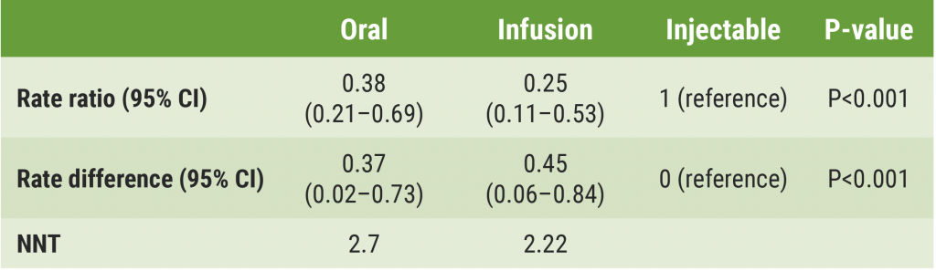
There was lower perfusion in white matter lesions than in normal-appearing white matter (0.43 vs 0.49; P<0.001). However, single-lesion R1 values were very heterogeneous (range 0.08-2.5). In single lesions, higher baseline perfusion was associated with more extensive remyelination (β=0.32; P<0.001) and reduced demyelination (β=-0.28; P<0.001). Lesion-specific perfusion at baseline was an independent predictor of successful myelin repair (OR 8.4; P<0.001).
- Colombi A, et al. Lesion-specific perfusion levels affect myelin loss and repair in multiple sclerosis: a positron emission tomography study. MSVirtual 2020, Abstract PS11.03.
Posted on
Table of Contents: MS Virtual 2020
Featured articles
COVID-19 and MS
Biomarkers
Treatment Strategies and Results
Management of progressive MS with approved DMT
Novel Treatment Directions
Neuromyelitis Optica Spectrum Disorders
Miscellaneous Topics
site created by:

© 2024 Medicom Medical Publishers. All rights reserved. Terms and Conditions | Privacy Policy