VDRF are common in MS, affecting over 50% of patients, with hyperlipidaemia, hypertension, and obesity being most prevalent [1]. Disease progression in MS is accelerated by the presence of VDRF, but the mechanisms behind this association are not well known. Dr Vijayshree Yadav (Oregon Health & Science University, USA) and her group hypothesised that VDRF leads to a reduced substrate delivery, which increases mitochondrial dysfunction. Subsequently, this could lead to increased high-energy phosphate metabolite deficiencies, increased neurodegeneration, and disease progression. Dr Yadav presented results from the first study using 7T MRS to longitudinally assess brain metabolism in MS subjects with and without VDFR [1].
The study included 52 patients with MS; 29 were VDRF-positive and 23 VDRF-negative at baseline. They were subjected to 7T MRS/MRI to assess differences in high-energy phosphate metabolites, brain volumes, and associated disease progression. Brain volumes were reduced in VDRF-negative patients. VDRF-positive patients had approximately 5% lower brain ATP than VDRF-negative patients (see Figure). The MRI signal changes were consistent in white and grey matter. Patients in the VDRF-positive group had higher neurological disability as assessed by EDSS.
Figure: Brain ATP is reduced in VDRF-positive MS patients [2]
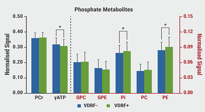
VDRF, vascular disease risk factors; PCr, phosphocreatine; ATP, adenosine triphosphate; GPC, glycerophosphocholine; GPC, glycerophosphoethanolamine; Pi, inorganic phosphate; PC, phosphocholine; PE, phosphoethanolamine.
Dr Yadav concluded that ATP reductions in VDRF-positive MS patients are possibly caused by lower cranial blood flow and are related to mitochondrial dysfunction, which could contribute to accelerated disease progression.
- Marrie R, et al. Neurology. 2010 Mar 30;74(13):1041-7.
- Yadav V, et al. Vascular Disease Risk Factors in Multiple Sclerosis (MS) is Associated with Brain Adenosine Triphosphate Abnormalities: Dysmetabolism May Drive MS Disease Progression. S2.004, AAN 2021 Virtual Congress, 17-22 April.
Copyright ©2021 Medicom Medical Publishers
Posted on
site created by:

© 2024 Medicom Medical Publishers. All rights reserved. Terms and Conditions | Privacy Policy
HEAD OFFICE
Laarderhoogtweg 25
1101 EB Amsterdam
The Netherlands
T: +31 85 4012 560
E: publishers@medicom-publishers.com